Our Portfolio
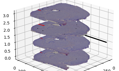
Created a 3D Python visualization of brain slices for a neuroimaging study
3D Images
Data Analysis and Visualization
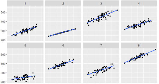
Generated output to determine Bayesian inference parameters
Bayesian Inference
Statistics
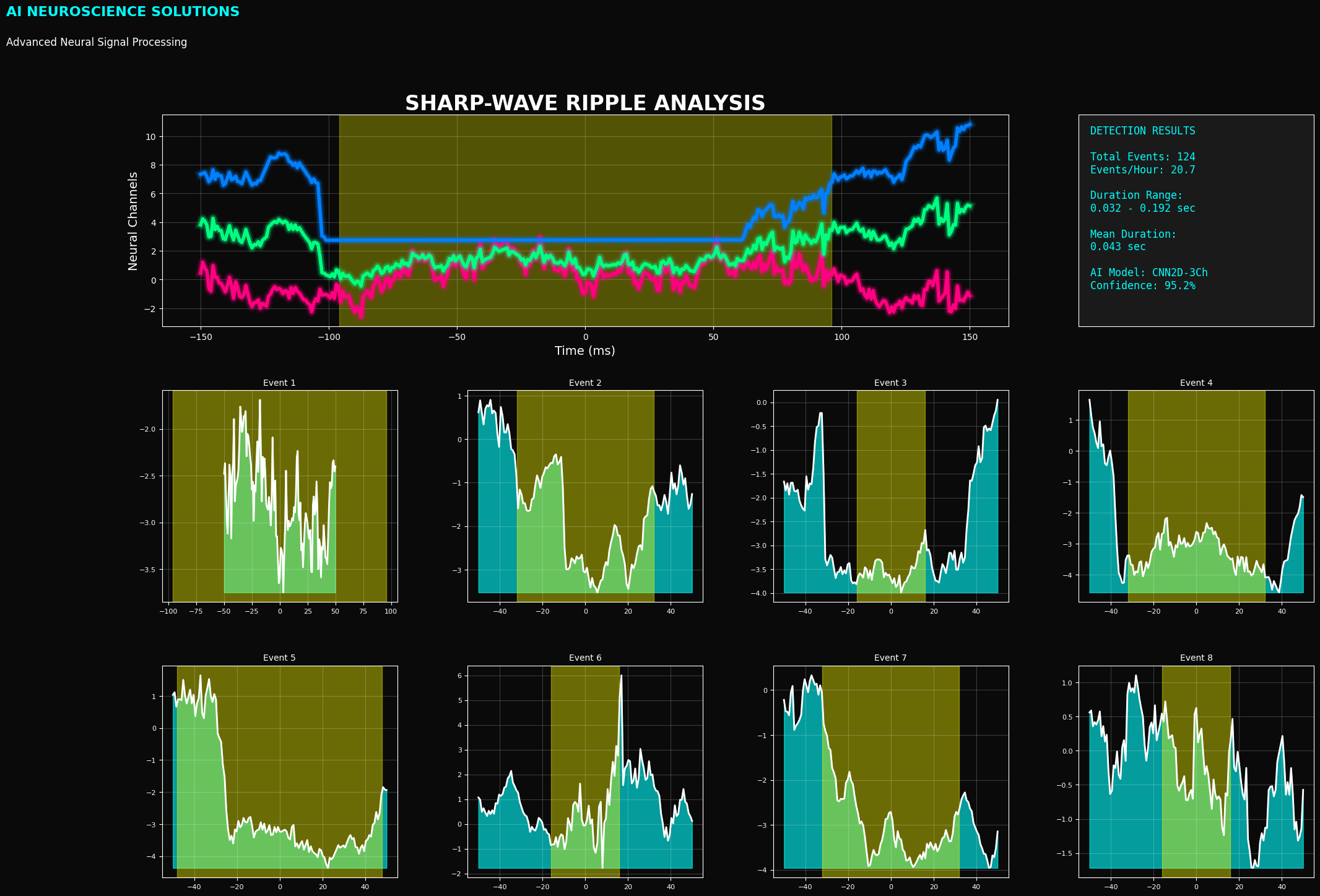
Developed a Python tool to visualize sharp-wave ripple detection
Sharp-Wave Ripples
Data Analysis and Visualization
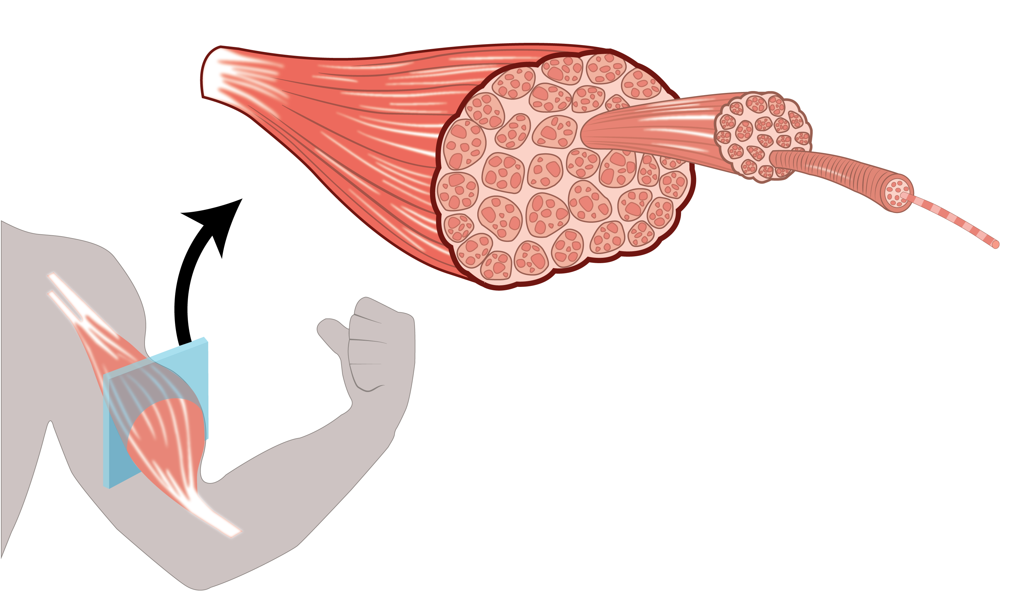
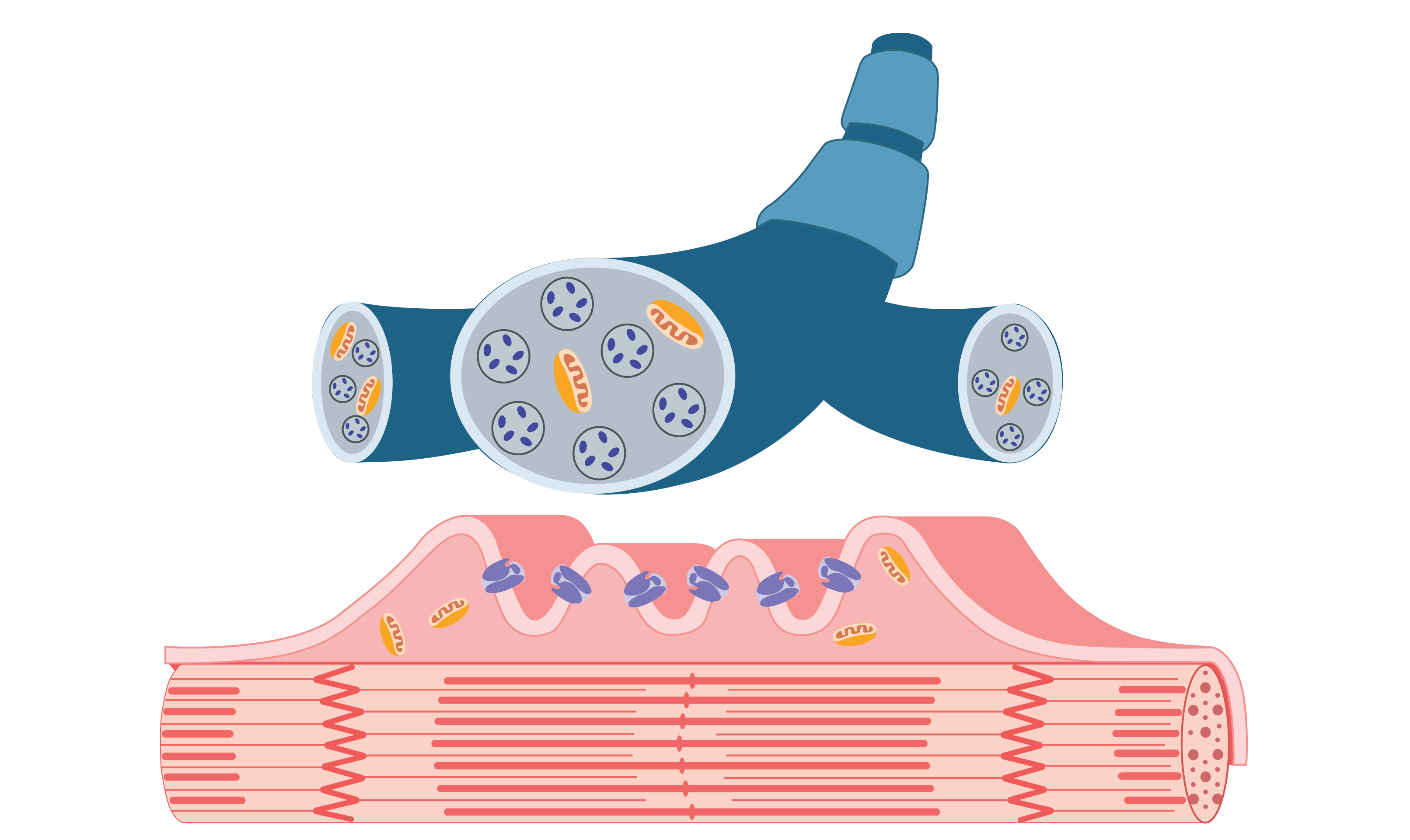
A cross-sectional view of a specific muscle, in this case, the biceps, with visible microstructures that serve as the basis for muscle contraction
Muscle Contraction
Biophysics
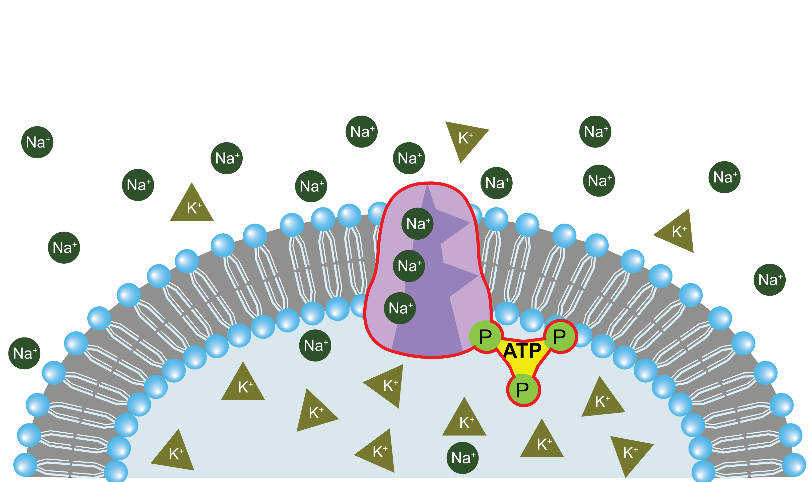
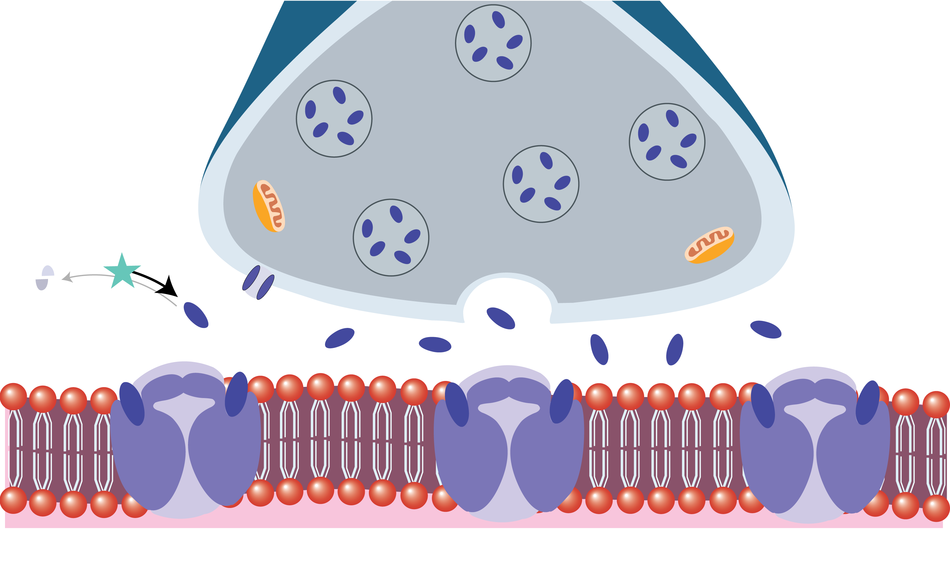
Illustration depicting a normal cell, highlighting the role of the sodium-potassium ATPase pump
Sodium-Potassium Pump
Cell Biology
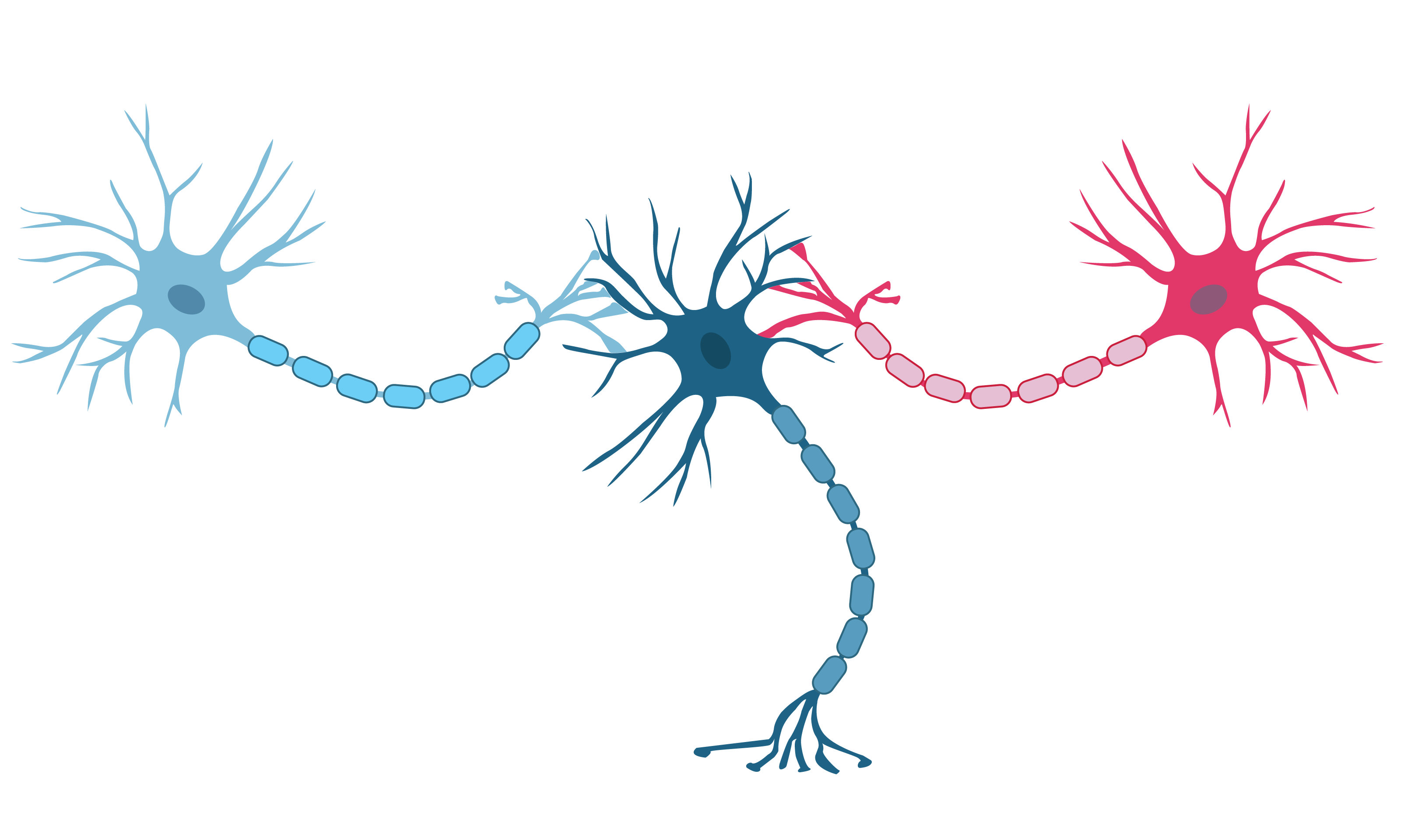
Synapses in the central nervous system, highlighting the release of neurotransmitters after the arrival of an action potential at the axon terminal
Synapses
Neurophysiology
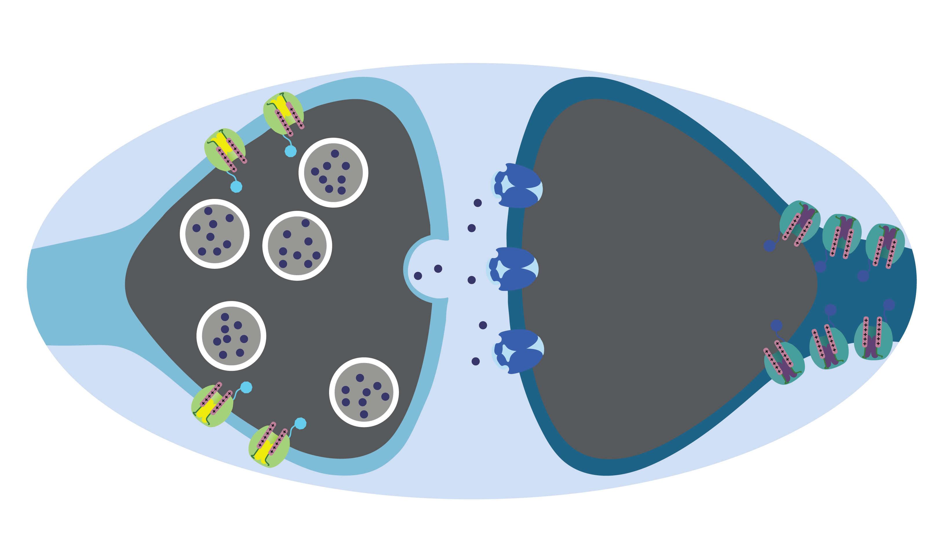
Multiple synapses acting on the post-synaptic cell, illustrating the concept that the activity of these cells is determined by the balance between excitatory and inhibitory inputs
Postsynaptic Cells
Neurophysiology
The neuromuscular junction receiving inputs from alpha motor neurons. When activated, these neurons trigger the release of acetylcholine, which sets off a signaling cascade in the muscle fiber, ultimately leading to muscle contraction
Muscle Contraction Trigger
Physiology
Neuromuscular junction, showing the release of acetylcholine and its eventual binding to acetylcholine receptors on the motor end plate of a muscle fiber
Neuromuscular junction
Cell Biology
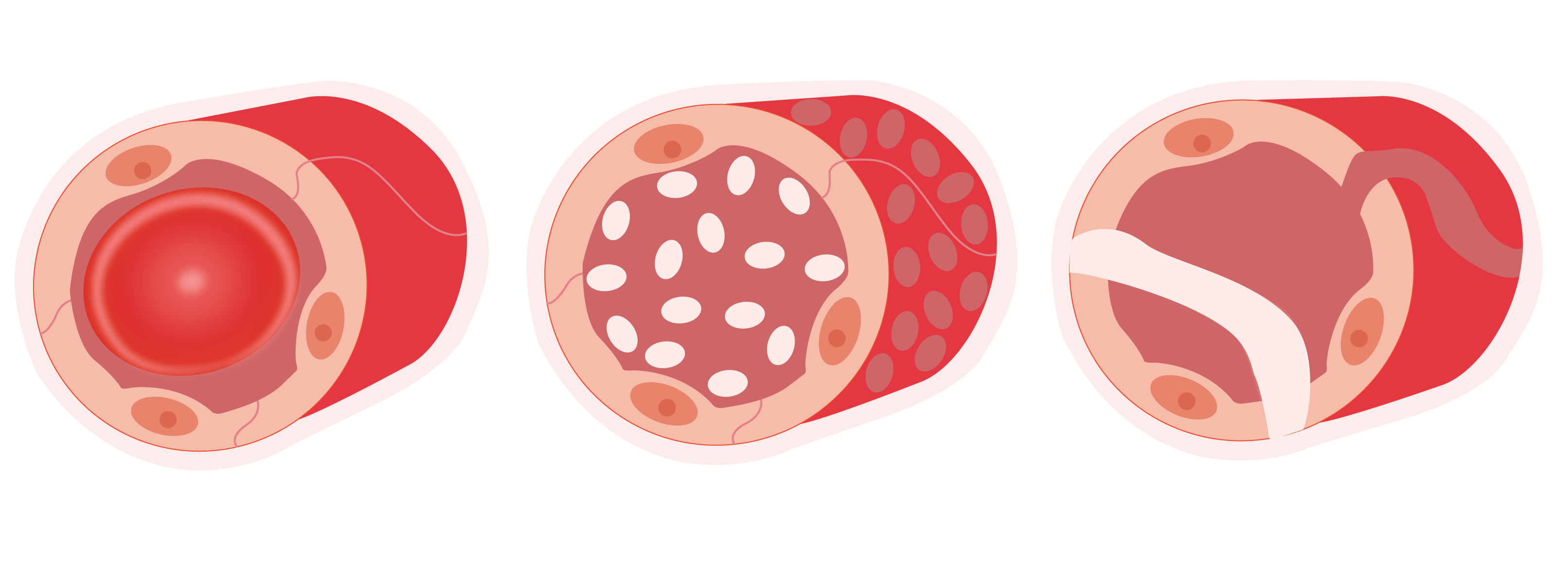
Illustration showing three different types of capillaries found in humans: continuous capillary, fenestrated capillary, sinusoidal capillary
Human Capillary System
Physiology
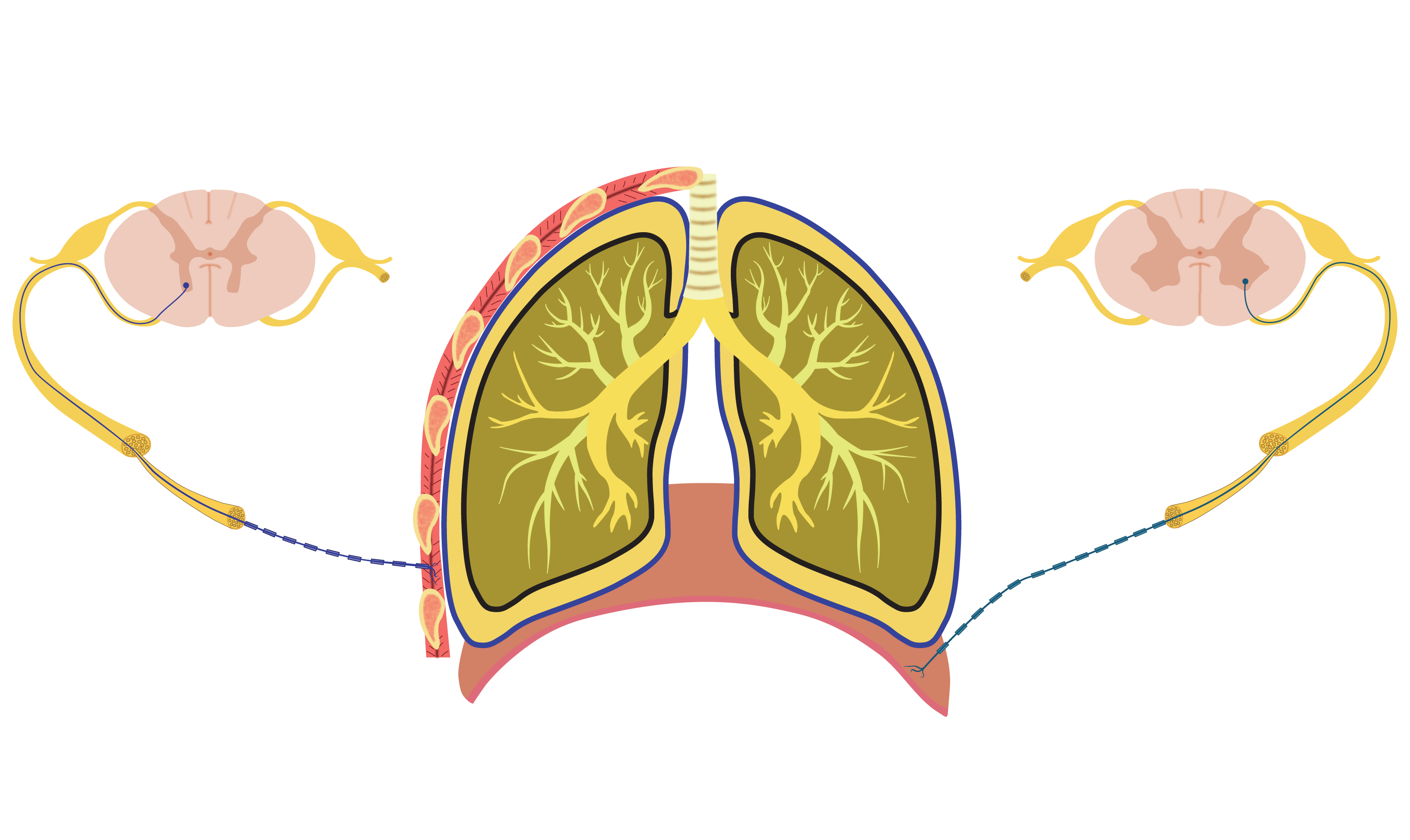
This image emphasizes the muscular innervation of the diaphragm and external intercostal muscles, the primary respiratory muscles involved in the normal respiratory cycle
Diaphragm Muscular Innervation
Physiology
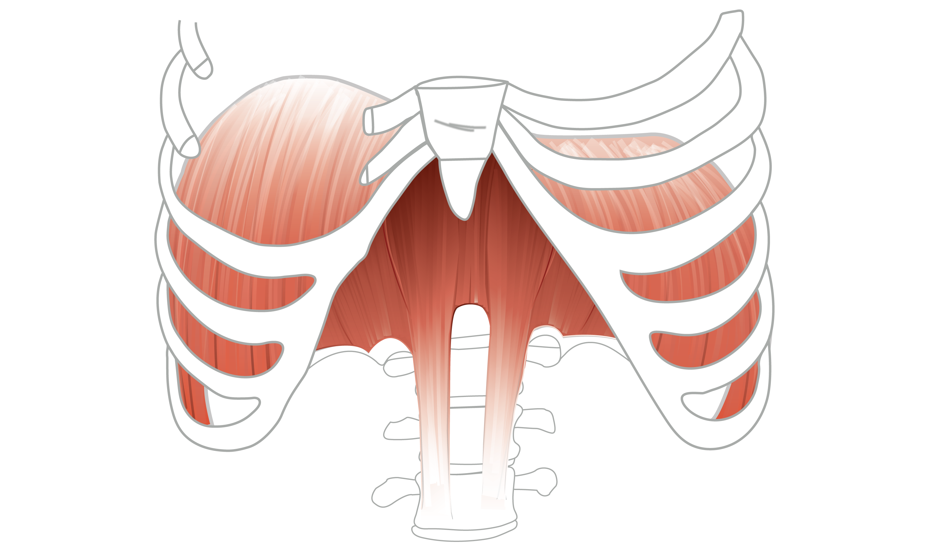
This image shows the thoracic cage, highlighting the role of the diaphragm, which is fully relaxed at the end of the respiratory phase
Thoracic cage
Anatomy
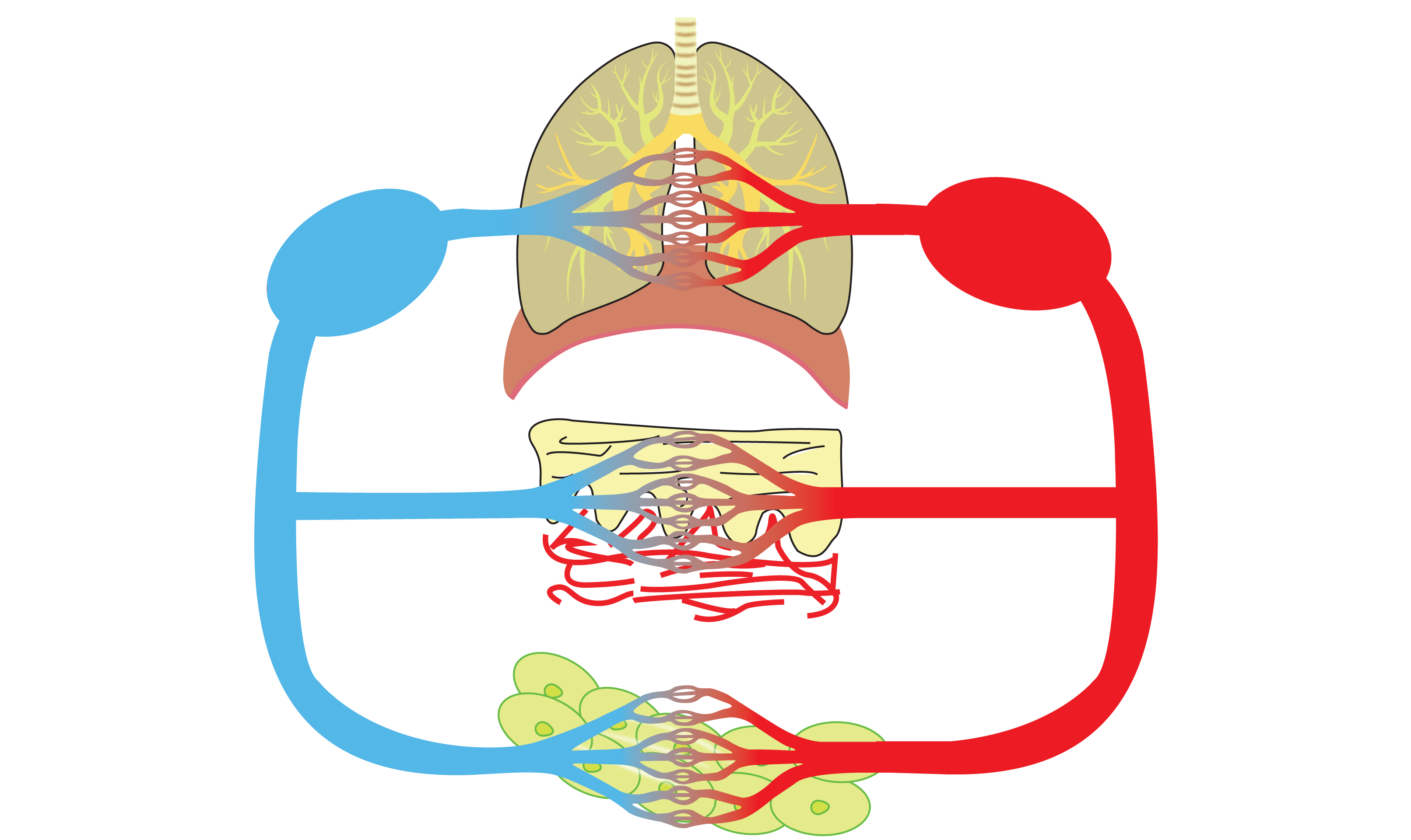
External and internal respiration in the pulmonary sustem. The oxygenated blood is conveyed to specific tissues, with the skin represented here, while the lower image depicts a generic body cell
Respiratory System
Physiology
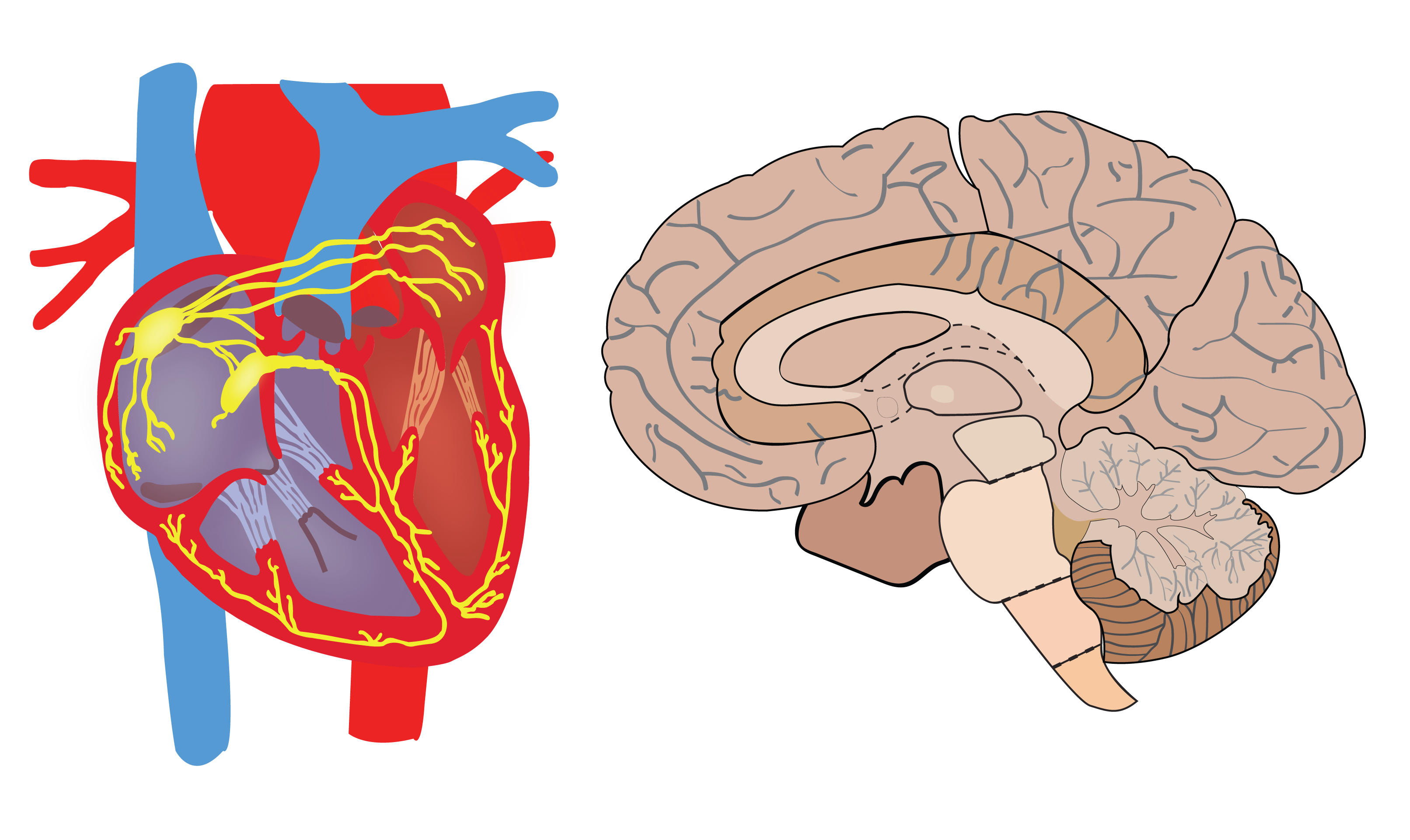
These images represent the heart and the brain, highlighting crucial structures for understanding their roles in physiology
Heart & Brain
Anatomy
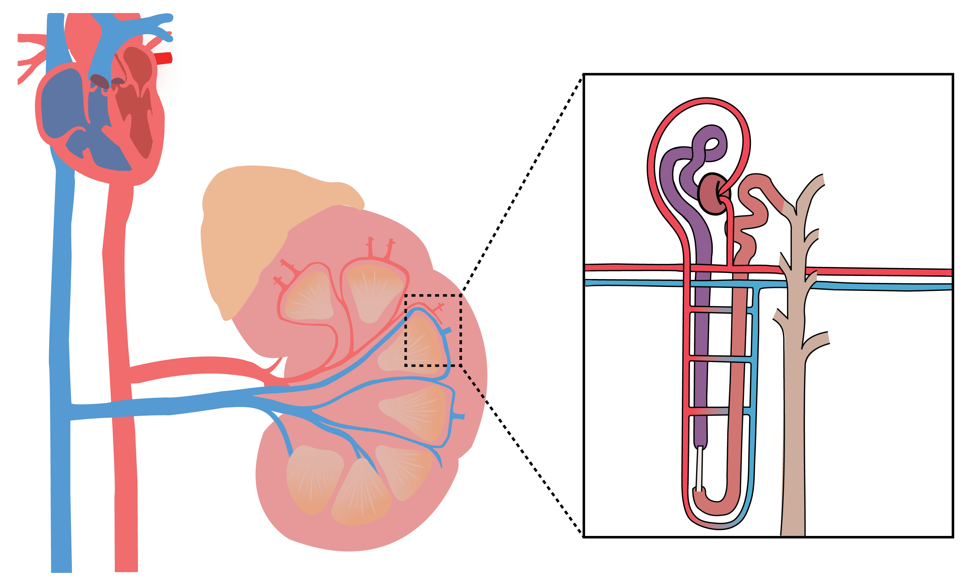
Magnified portion of the kidneys, specifically the cortex, where individual nephrons are visible, forming the base for urine formation
Kidney & Nephron
Physiology
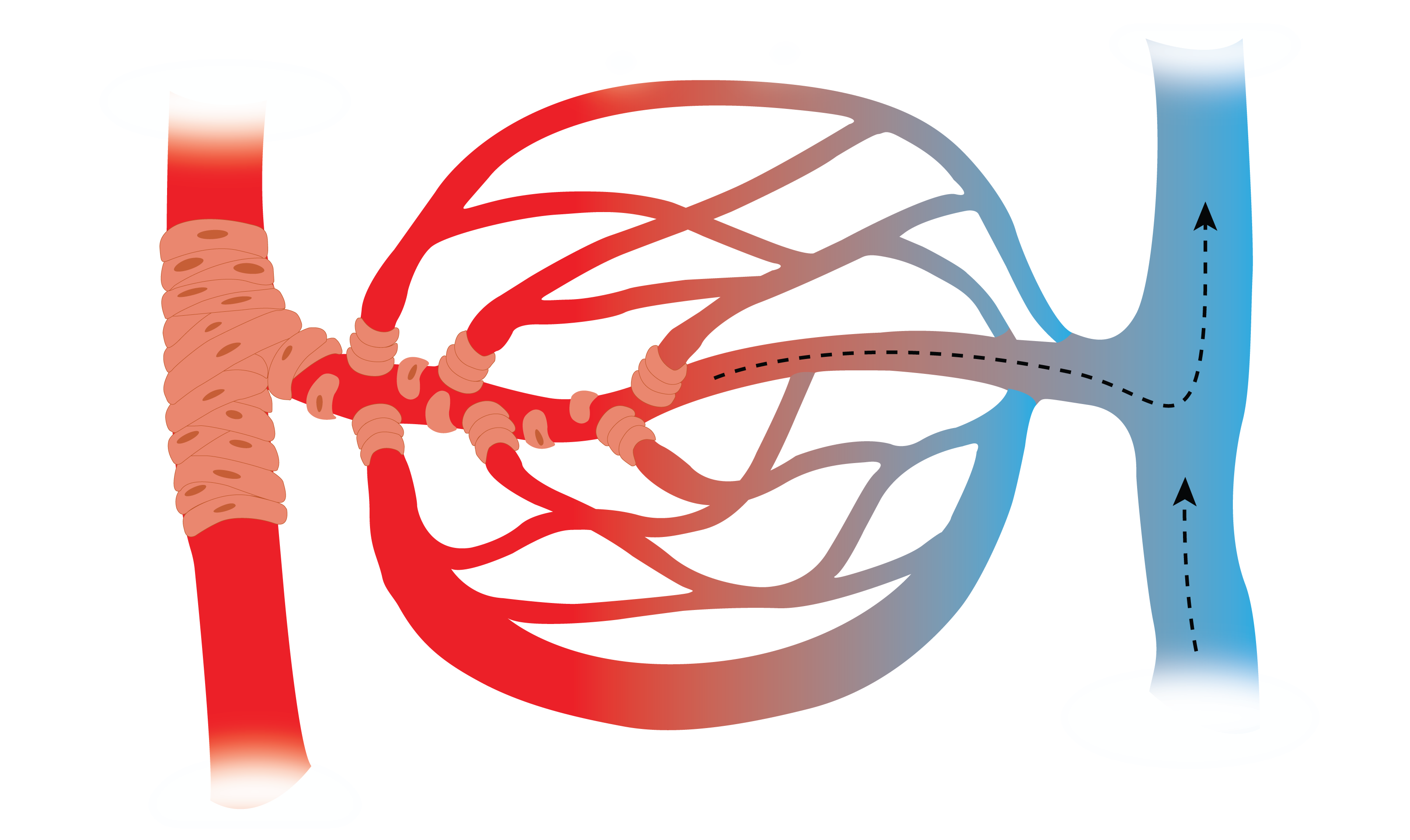
Illustrated capillary beds to highlight gas and nutrient exchange
Circulatory System
Physiology
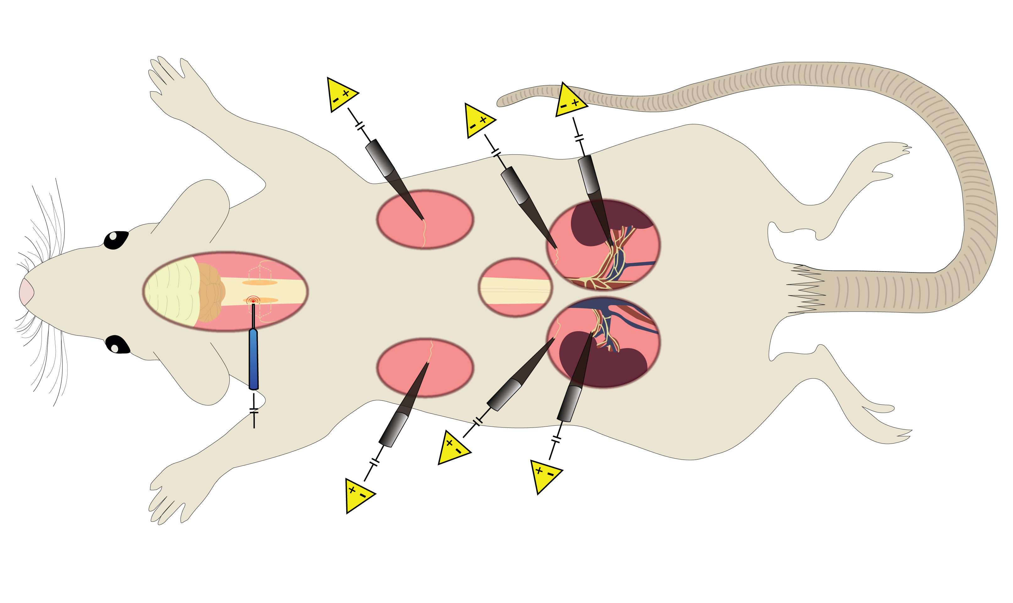
A mouse used in neuro surgeries, with electrodes are implanted for recording local field potentials and muscle activity. Electrode placements in the spinal cord, muscles, and brain regions
Neuro Surgeries
Neuroscience
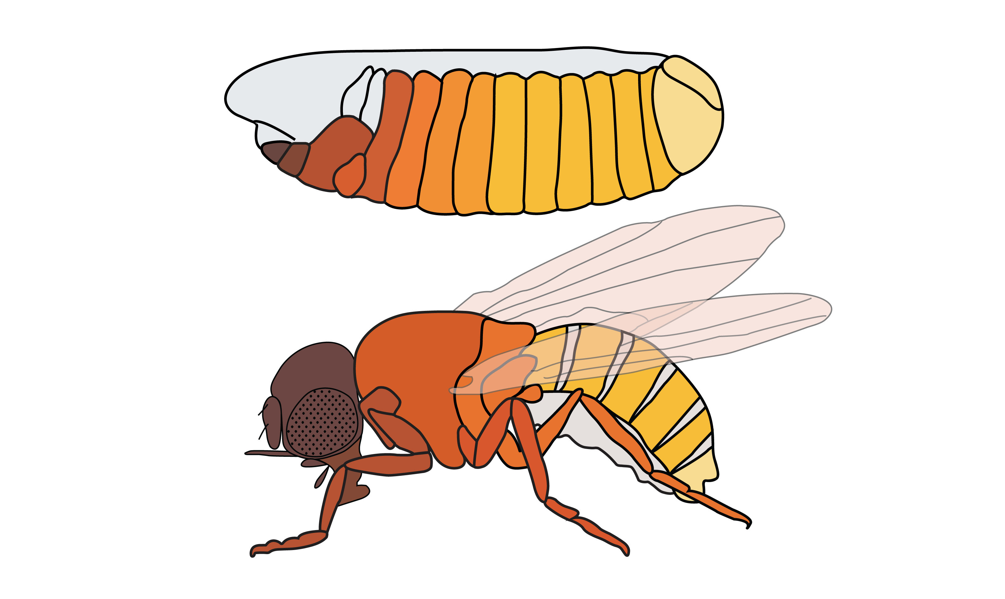
This image is a visual representation of biological development, particularly in the context of the HOX gene, which plays a crucial role in regulating the development of body structures in organisms
Drosophila
Morphology